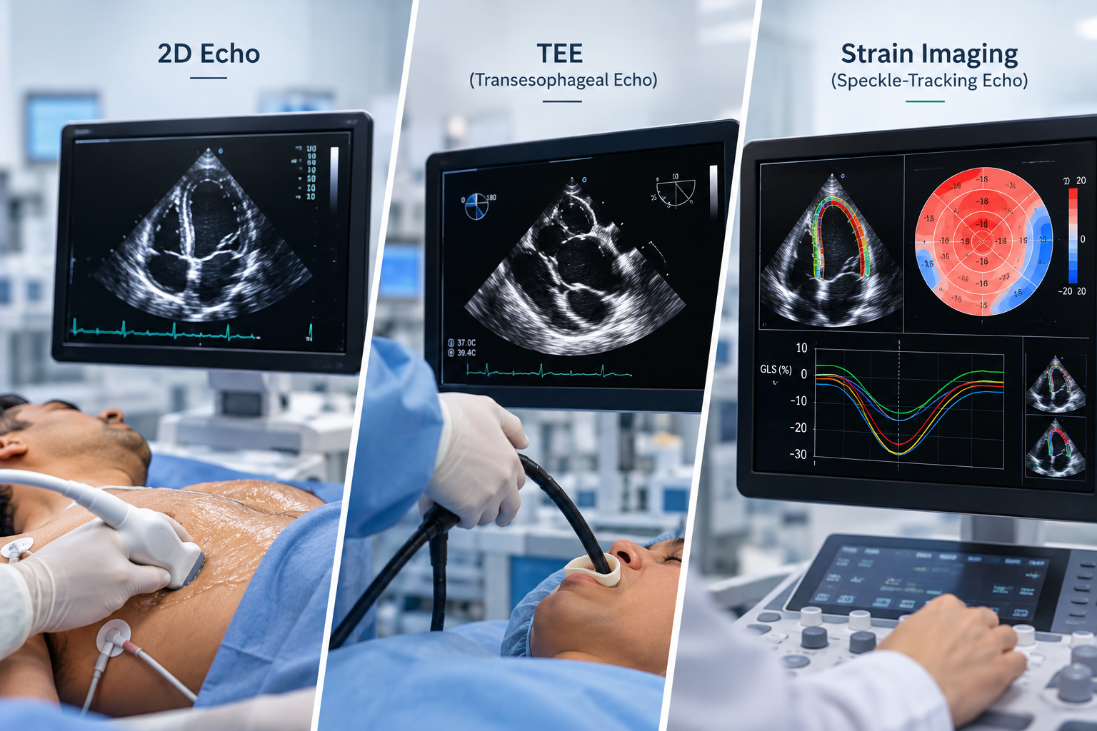
Detailed Imaging of Heart Structure and Function
Echocardiography uses ultrasound technology to evaluate the structure and functioning of the heart. Dr. Rohan Ainchwar provides access to advanced cardiac imaging including 2D Echocardiography, Transoesophageal Echocardiography (TEE) and Strain Imaging, according to clinical requirements.
The available 2D echocardiography services include adult, pediatric and fetal echocardiography, supporting cardiac assessment across different patient groups.
2D Echocardiography
A 2D Echo produces real-time images of the heart and allows assessment of its chambers, valves and pumping function. It can provide important information when evaluating conditions such as heart failure, valvular heart disease, cardiomyopathy and other structural cardiac abnormalities.
Pediatric and Fetal Echocardiography
Pediatric echocardiography is used to evaluate heart structure and function in children. Fetal echocardiography focuses on the developing baby’s heart during pregnancy when detailed cardiac assessment is clinically indicated.
Transoesophageal Echocardiography (TEE)
In TEE, a specialized ultrasound probe is passed into the oesophagus, allowing certain cardiac structures to be viewed from a closer position than with conventional echocardiography.
TEE may provide additional information when detailed visualization of the heart valves, chambers or other structures is required.
Strain Imaging
Strain imaging is an advanced echocardiographic technique that evaluates myocardial deformation and provides additional information about the functioning of the heart muscle.
Echocardiography May Help Assess
- Heart pumping function
- Heart valve abnormalities
- Structural heart conditions
- Cardiomyopathy
- Selected congenital abnormalities
- Changes in heart muscle function
The specific echocardiographic technique recommended depends on the patient’s symptoms, medical history and findings from the initial cardiac evaluation.